Tag Archives: Knees

Studies: Osteoarthritis Patients Using Exercise Therapy Have Less Pain, Cut Opioid & Analgesic Use

Conclusion Among patients with knee or hip OA using analgesics, more than half either discontinued analgesic use or shifted to lower risk analgesics following an 8-week structured exercise therapy and patient education programme (GLA:D). These data encourage randomised controlled trial evaluation of whether supervised exercise therapy, combined with patient education, can reduce analgesic use, including opioids, among patients with knee and hip OA pain.

Read study

Health Study: Physical Therapy Superior To Steroid Injection For Knee Osteoarthritis

New England Journal of Medicine Articles

Patients with osteoarthritis of the knee who underwent physical therapy had less pain and functional disability at 1 year than patients who received an intraarticular glucocorticoid injection.

Physical Therapy Superior To Glucocorticoid Injection for Knee Osteoarthritis NEJM April 9 2020

Osteoarthritis of the knee is a leading cause of disability.1 Current management is typically limited to the treatment of symptoms until late stages of arthritis lead to knee replacement.2 Intraarticular glucocorticoid injections are commonly used as a primary treatment for osteoarthritis of the knee,3 but there are conflicting reports regarding the extent and duration of the relief of symptoms with this therapy.4-6 Complications from these injections occur infrequently but include joint infection,7 accelerated degradation of articular cartilage,8 and subchondral insufficiency fractures.9

PHYSICAL THERAPY

The physical therapy intervention, which is described in the protocol,26 included instructions and images for exercises, joint mobilizations, and the clinical reasoning underlying the priorities, dosing, and progression of treatment. During a typical clinical session, the physical therapist would implement hands-on, manual techniques immediately before the patient performed reinforcing exercises to help the patient perform the movements with little or no pain. For example, if a patient could not fully extend or flex the knee, or those movements were painful, the physical therapist would use a hands-on, passive mobilizing technique to repeatedly move the knee to reduce stiffness while altering the mechanics of the technique to avoid pain. The patient would then perform repeated active knee movements in the same direction.

Read full study

Study: Middle-Aged Knees With Meniscal Tears – No Benefits To Surgery Over Exercise Therapy (BMJ)

Exercise or Surgery - Meniscal Tear in Middle-Aged Knees BMJ

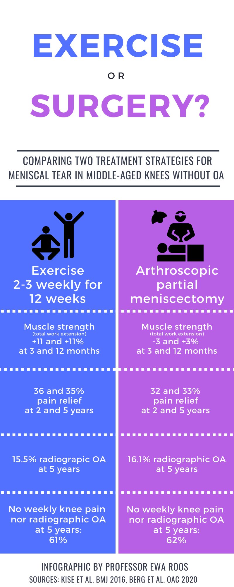
Conclusion: The study was inconclusive with respect to potential differences in progression of individual radiographic features after surgical and non-surgical treatment for degenerative meniscal tear. Further, we found no strong evidence in support of differences in development of incident radiographic knee osteoarthritis or patient-reported outcomes between exercise therapy and arthroscopic partial meniscectomy.

Objective: To evaluate progression of individual radiographic features 5 years following exercise therapy or arthroscopic partial meniscectomy as treatment for degenerative meniscal tear.

Design: Randomized controlled trial including 140 adults, aged 35-60 years, with a magnetic resonance image verified degenerative meniscal tear, and 96% without definite radiographic knee osteoarthritis. Participants were randomized to either 12-weeks of supervised exercise therapy or arthroscopic partial meniscectomy. The primary outcome was between-group difference in progression of tibiofemoral joint space narrowing and marginal osteophytes at 5 years, assessed semi-quantitatively by the OARSI atlas. Secondary outcomes included incidence of radiographic knee osteoarthritis and symptomatic knee osteoarthritis, medial tibiofemoral fixed joint space width (quantitatively assessed), and patient-reported outcome measures. Statistical analyses were performed using a full analysis set. Per protocol and as treated analysis were also performed.

Results: The risk ratios (95% CI) for progression of semi-quantitatively assessed joint space narrowing and medial and lateral osteophytes for the surgery group were 0.89 (0.55-1.44), 1.15 (0.79-1.68) and 0.77 (0.42-1.42), respectively, compared to the exercise therapy group. In secondary outcomes (full-set analysis) no statistically significant between-group differences were found.

Read full study